Case Study
Incidental Finding Not Followed
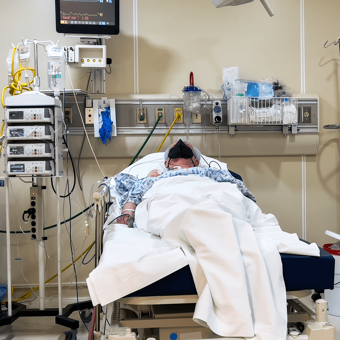
Description
A 42-year-old woman was diagnosed with lung cancer three years after a chest X-ray—done to locate a swallowed dental crown—revealed a mass in her upper right lobe.
Key Lessons
- A reliable system to reconcile and communicate serious unrelated findings from imaging studies is essential to safe, high-quality care.
- Patients who are made aware of abnormal findings can help ensure necessary follow-up with their providers.
- A non-specific finding and inconclusive mammogram in the presence of a palpable mass requires further workup with ultrasound.
- Patients and juries may expect that the ordering provider will fully review the resulting imaging reports and follow up on all of its significant findings.
Clinical Sequence
About three weeks after swallowing a dental crown, a 39-year-old woman went to a thoracic surgeon who ordered a thorax CT scan, which confirmed that she had aspirated a foreign object. The radiology report also included an (incidental) finding of a 1cm x 6mm nodule in the patient’s right upper lobe; follow up with a CT scan within three months was recommended. The report was posted immediately in the hospital’s computerized system. There is no documentation of any direct communication between Radiology and the surgeon. The primary care physician did not receive a copy of the report.
Two days after the CT scan, a bronchoscopy was performed to remove the dental crown. During the procedure, the surgeon and Radiology discussed the CT scan, but it was not available in the OR. The patient did not return to the surgeon for recommended follow-up. The surgeon did not initiate follow up with her about the lung nodule.
Nearly three years later, a chest X-ray prompted by persistent shoulder pain showed a right apical mass. The patient was subsequently diagnosed with lung cancer. She expressed anger and sadness upon learning of the earlier CT finding of a suspicious mass, and requested a change of PCPs. Following two years of aggressive treatment, the patient experienced regional recurrence suggestive of metastatic disease, with a grim prognosis.
Allegation
The patient sued the thoracic surgeon, two radiologists, and the anesthesiologist, alleging a three-year delay in diagnosis of lung cancer.
Disposition
The case was settled in the high range (>$500,000)
Analysis
The surgeon who ordered the CT scan and learned that Radiology found the dental cap in the upper lobe bronchus does not recall how the information came to him, but suggests “it could have come from his secretary, a radiologist, or a resident.” The radiologist recalls calling the surgeon’s office with the incidental findings two days before the surgery, but doesn’t know who he talked to. Neither communication was documented.
Protocols for all involved specialty services can help ensure that the responsible provider is aware of important findings in a timely way. A surgeon hearing the findings second-hand may not fully appreciate the severity of the results. Likewise, a radiologist who does not know to whom the information is being given, cannot be assured that the responsible provider is getting all the pertinent information.
The final radiology report was transcribed the day after the surgeon bronchoscoped the patient to remove the object. The scans had been posted immediately in the hospital computer system, but the terminals in the surgeon’s office and in the OR could not access them. The surgeon’s routine practice was to go to the Radiology department before surgery to view the films—it is not clear if he did so for this case. The radiologist recalls getting a phone call from the OR to discuss the films. The surgeon never received a written copy of the CT scan report.
Ordering physicians need a system that tracks reports that they have ordered. The ordering physician is responsible for reconciling all ordered tests and for executing follow up of all abnormal results—including incidental findings. The incidental finding in this report of a suspicious mass in the upper right lobe was important. If the surgeon had been aware of the unexpected lung mass, a follow-up CT scan three months later as recommended by the radiologist may have been pursued and improved the patient’s outcome.
Following surgery, the patient was told to return to the surgeon’s office for a post-operative follow-up. The patient never returned; her medical record has no record of phone calls to or from the patient after the surgery. In addition, the surgeon never pursued the recommended follow-up CT.
Reminders to patients can be helpful in ensuring that needed postoperative follow up occurs. A patient who does not call when recommended may not fully understand the significance or the need. This can be addressed ahead of time with explicit explanations about the reasons follow-up is important. Putting simple instructions in writing is more effective than oral instructions alone. A “tickler system” in the physician’s office can identify who is due for follow up and prompt a call or note to the patient from the office.
The patient was mad at the surgeon for not returning two phone calls from her after the bronchoscopy.
Unreturned calls can lead patients to believe the physician is disorganized or doesn’t care. Practices with very clear guidelines in place for who should handle patient telephone calls and how they should be managed can avoid such perceptions. An office practice system that captures every patient call and tracks the needed follow up can support good care and defend it.
Patients, more than anyone else, need to know what their providers know about their health.
If the patient had been included in the trail of information about her CT results, she may have been able to play an active role in ensuring appropriate follow-up. She expected the test would find the aspirated cap, but had no indication of an incidental, worrisome finding.
Perioperative notes by the anesthesiologist and the surgeon reference the CT finding of the aspirated dental cap, but not the incidental finding of a suspicious lung mass. The surgeon did not remember how he found out about the CT results. The radiologists recalled telling someone in the surgeon’s office about the incidental finding of a nodule, but did not know whom she spoke with.
Physicians should document all basic facts surrounding their encounters with one another. Also, making contemporaneous notes about what was communicated to the patient is critical. Documenting when, how, and to whom the findings are communicated can help identify where systems break down and how to understand what went wrong. Such documentation can also be helpful in defending against a claim of negligence.
A review of the practices in this hospital revealed that this was not an isolated problem. Communication of critical test results, especially incidental findings, was found to be unreliable.
Remedially, the hospital developed protocols and guidelines that would help ensure a reliable system for giving and receiving important study findings for every patient in a timely way.
The presence of a suspicious mass identified on a CT scan with no follow-up or discussion with the patient for two years, along with inadequate documentation—and dispute amongst the defendant clinicians—about what was communicated—merited settlement of this case.
Finger-pointing and blame are serious problems for the defense of a professional liability claim. Patients and juries who see evidence of that tend to view the blame as applying to every defendant when they see that. Documentation can help a provider show that his or her actions were appropriate at all times.
This is a fictitious case that illustrates commonly encountered issues and is for educational purposes only. Any resemblance to real persons, living or dead, is purely coincidental.
See More MPL Cases
Medication Mix-up Contributes to Patient’s Death

A Devastating HIT: When a Delayed Diagnosis Costs Limbs

Retaliation Allegation after Mental Health Leave